WelcomeSee more
“
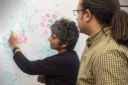
One of the hallmarks of the Department of Neuroscience is the level of interaction and collaboration between laboratories and laboratories in other departments.
”
RICHARD HUGANIR, PhD, Director

Faculty MembersSee more
-
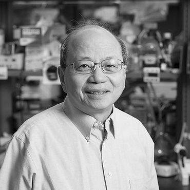
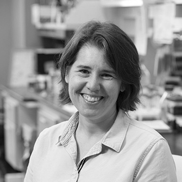
 Jay Baraban MD, PhD Professor of Neuroscience
 -
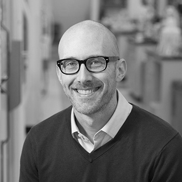
 “We want to understand our learning and cognition by cells and synapses.” Hyungbae Kwon PhD Associate Professor of Neuroscience
Director of Admissions -
 “Our laboratory studies phototransduction and olfactory transduction.” King-Wai Yau PhD Professor of Neuroscience
 -
 Ulrich Mueller PhD Bloomberg Distinguished Professor of Neuroscience and Biology
 -
 “My research interest is in understanding how sensory experience alters brain circuits.” Hey-Kyoung Lee PhD Professor of Neuroscience
 -
 “We study neural circuits for sensory perception, with a focus on touch.” Daniel O'Connor PhD Professor of Neuroscience
Co-Director of the Neuroscience Training Program -
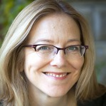
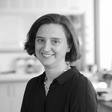 “My lab studies how brain circuits process sensory information to generate perception.” Solange Brown MD, PhD Associate Professor of Neuroscience
 -
 “We study how the auditory periphery develops and its ability to regenerate.” Angelika Doetzlhofer PhD Associate Professor of Neuroscience
EventsSee events calendar
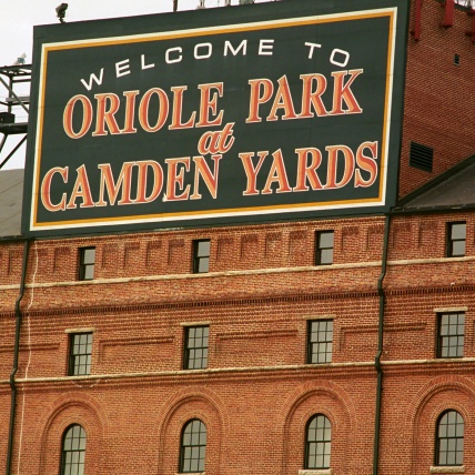
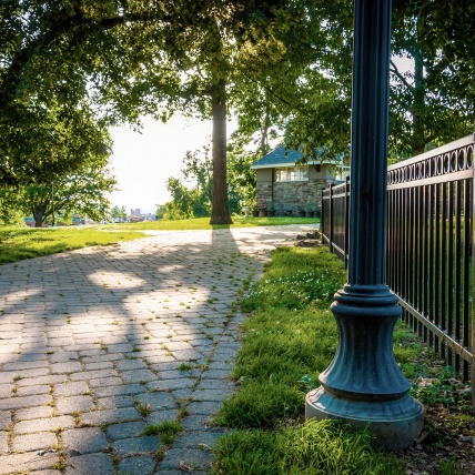
Baltimore citySee more
-
“ The waterfront promenade is a great way to enjoy a sunny day in Baltimore. From Harborplace, you can walk east through lovely waterfront neighborhoods—Harbor East, Fells Point,Canton. Great watering holes, pubs, and coffee shops! ” Ronald Schnaar, Professor“ The Chesapeake Bay is one of the premier sailing areas in the world. I love sailing my boat and spending time on the water. ” Ernst Niebur, Professor“ I love the Crab Cake Cones from the Gypsy Queen (gypsyqueencafe.com) food truck when they’re on campus. ” Jenn Orthmann Murphy, MD, PhD (Postdoc, Bergles Lab)
-


-
“ The year-round ultimate frisbee leagues in Catonsville and the Neuroscience department softball team. ” Travis Faust, Graduate Student (Sawa Lab)“ I’m new here, so getting lost biking through Baltimore has been a great way to explore. The murals are beautiful. Also, it’s wonderful to be living in a city that is celebrated for their crab dishes—my favorite comfort food! ” Raina D’Aleo, Graduate Student“ Each neighborhood in Baltimore has a unique personality offering a variety of affordable food options. I love the charcuterie selection at Trinacria and Parts & Labor. ” Wendy Xin, Graduate Student (Bonci Lab)