Xinzhong Dong PhD
Professor of Neuroscience, Neurosurgery, Dermatology
Professor of Neuroscience, Neurosurgery, Dermatology

Biochemistry, Cellular and Molecular Biology Graduate Program
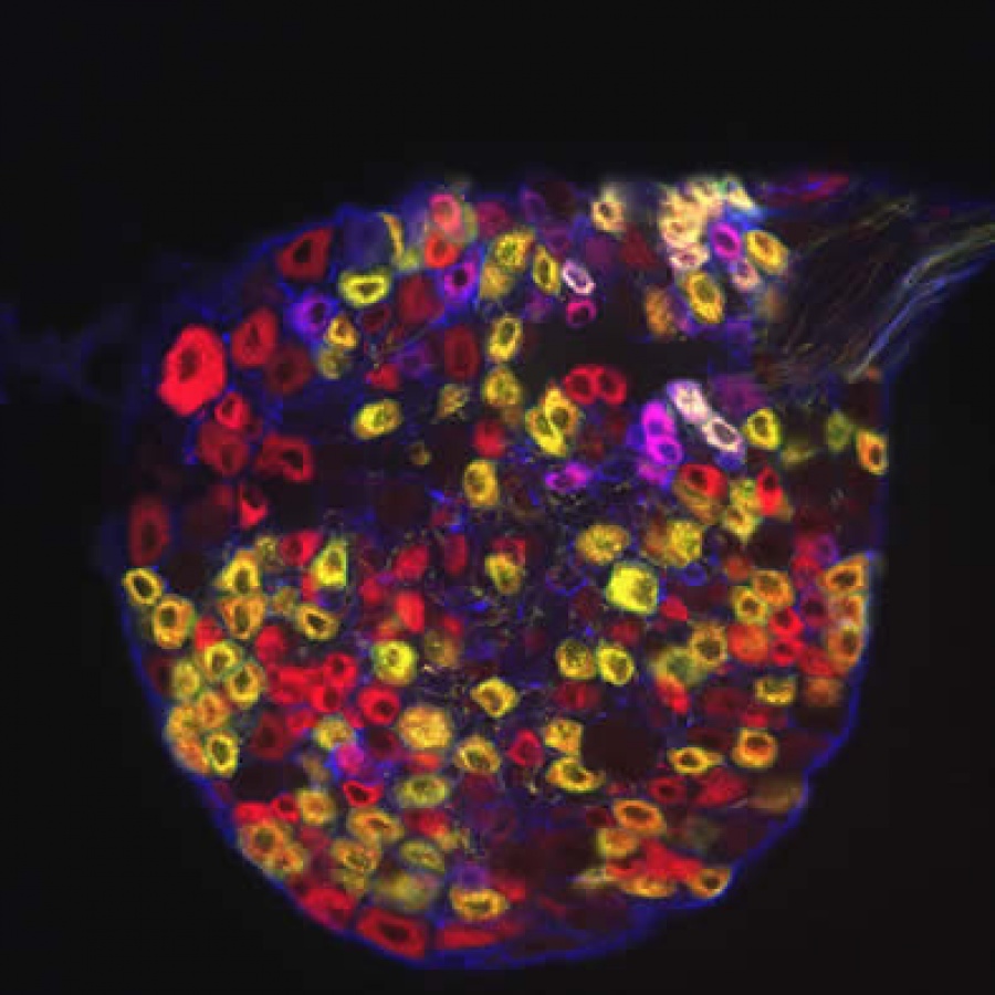
 The DRG section was immunostained with three different antibodies to visualize MrgD, MrgB4, and c-Ret positive neurons. This data indicates high degree of molecular diversity in DRG neurons.

The DRG section was immunostained with three different antibodies to visualize MrgD, MrgB4, and c-Ret positive neurons. This data indicates high degree of molecular diversity in DRG neurons.
Pain is required for survival of the organism. However, chronic pain resulting from inflammation and nerve injury can seriously undermine the quality of life. Pain or nociception is mainly mediated by a subset of primary sensory neurons, known as nociceptors, in dorsal root ganglia (DRG). Our research goal is to understand molecular mechanism of pain. To achieve this goal, I started with searching mouse genes specifically expressed in nociceptors. From a very unique screen technique, I identified a large family of G protein-coupled receptor (GPCRs), called Mrgs, comprising nearly 50 members. Some of Mrgs were only found in nociceptors but not in any other tissues in the body. Interestingly, the expression of different Mrgs is largely non-overlapping in the nociceptors, suggesting that there is an unexpectedly high degree of molecular diversity among these neurons. Using mouse molecular and genetic techniques, we found that the axons of Mrg expressing nociceptors mainly innervate skin but not visceral organs. To our knowledge, this is the first marker with such a specific innervation pattern. We also isolated several neuropeptides including FMRFamide, NPFF, and g2-MSH, function as ligands for Mrgs. Some of these Mrg ligands have been implicated in regulating pain sensitivity. Furthermore, our electrophysiological data indicated that activation of Mrg by the ligand could significantly increase the excitability of Mrg-expressing nociceptors. Together these results suggested that Mrgs maybe involved in chronic pain by sensitizing nociceptors. In my laboratory, I would like to extend the studies of Mrg function in pain. My recent data shows that the endogenous ligand for Mrgs is likely expressed by skin mast cells, which are in direct contact with Mrg-expressing nerve endings in the skin. We will employ biochemical methods to purify the ligand from mouse skin mast cells and test its ability to activate Mrgs and induce pain behaviors. To study Mrg function in vivo, we will generate several mouse lines in which a single Mrg gene or Mrg gene cluster will be deleted. We will determine whether these Mrg-deficient mice have any abnormalities in pain behaviors. Besides characterizing Mrgs, we will use recently developed genetic and molecular tools to study the cellular properties of Mrg-expressing nociceptors with respect to neuronal circuitry in central projection and pain modalities. GPCRs have been frequently used as drug targets for a variety of pharmacotherapies. Therefore, identification and functional analysis of Mrgs should open a door for the development of novel analgesics with limited side effects. Besides Mrg genes, I also isolated several novel nociceptor-specific molecules from the initial screen. Some of them encode ion channels and have very specific expression pattern similar to Mrgs. We will take the similar approaches as we did for the studies of Mrgs to characterize the functions of these genes in nociception. Our studies have shown that nociceptors are highly diverse at molecular level, and the roles of these neurons in nociception are far from understood. With nociceptor-specific genes in hand, we are in an advantageous position to combine molecular biology and genetics with animal behavior and electrophysiology to gain new insights into nociception.
Small-diameter nociceptive neurons whose cell bodies reside in dorsal root ganglia (DRG) and trigeminal ganglia (TG) play essential roles in pain signal detection, transmission, and modulation. The peripheral axons of these pseudo-unipolar neurons innervate skin, muscle, and visceral organs to detect painful stimuli, while their central axons transmit these signals to the spinal cord dorsal horn. One major contributor to hyperalgesia is augmented sensitivity of primary nociceptive neurons. Therefore, monitoring the activities of these neurons and axons is crucial to understand pain mechanisms. Imaging neuronal activity in primary sensory neurons from tissue explants and slices has been a challenge given that their nerve endings and cell bodies are densely surrounded by other cells (e.g. keratinocytes in the skin, glia surrounding somas, intrinsic neurons in the spinal cord) and therefore difficult to selectively load with Ca2+ sensitive chemical dyes. By specifically expressing a genetically-encoded Ca2+ sensitive indicator in almost all DRG and TG neurons in Pirt-GCaMP3 mice, we successfully detected activation of primary sensory neurons in the trigeminal system at peripheral and central terminals as well as in their cell bodies, in tissue explant and slice preparation. The advantages of this method are manifold: simple tissue preparation and imaging procedures; excellent spatial resolution; sensitive, high-efficiency simultaneous imaging of multiple neurons; preservation of somatotopic organization; and stable expression of GCaMP3 after nerve injury. Because of these advantages, we were able to visualize peripheral neuronal hypersensitivity in both injured and uninjured nerves corresponding to chronic pain from skin territories innervated by these nerves.